


The integration of artificial intelligence (AI) into healthcare has seen a remarkable surge in interest, particularly in its potential applications within the medical field. AI technologies promise to enhance processes such as swiftly analyzing samples or detecting disease markers that could elude human observers. However, a recent study suggests that the application of AI, specifically a method called virtual staining, may not always yield the expected benefits.
Research conducted by the Center for Label-free Imaging and Multiscale Biophotonics (CLIMB) indicates that while virtual staining can enhance the utility of medical images in certain scenarios, it may also hinder the acquisition of valuable information in others. Sourya Sengupta, a graduate student at the Beckman Institute for Advanced Science and Technology and the lead author of the study, emphasizes the need for caution in choosing whether to incorporate AI into specific workflows. “The general conclusion is that AI can be a great tool — it does help in some cases — but you have to be a little bit cautious,” Sengupta stated.
Understanding Virtual Staining
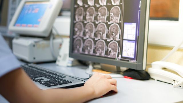
Most individuals have undergone medical imaging, whether through ultrasounds, MRIs, or X-rays, which are essential for diagnosing diseases and monitoring health. A specialized category of medical imaging involves microscopy images, which allow for detailed analysis of tissue and cellular samples. Traditionally, these samples are enhanced using staining techniques involving dyes or chemicals to improve contrast. Although effective, these methods can be time-consuming and potentially damaging to cells.
Label-free imaging presents an alternative to staining, utilizing the intrinsic properties of biological materials to generate images without chemical interference. This technique, however, often results in images with lower contrast, making it challenging to identify critical features. To address this limitation, researchers have turned to virtual staining, which employs computational models to predict how a label-free image would appear if it were stained.
The goal of virtual staining is to produce high-contrast images comparable to traditional staining but with the added benefits of speed and the preservation of sample integrity. Yet, it is crucial to validate the accuracy and practical utility of these virtually stained images for both clinical and biological explorations.
Methodology and Findings
The researchers evaluated the performance of virtually stained images against label-free and stained images across two specific tasks. The first task involved image segmentation, where a neural network identifies and isolates individual cell nuclei. The second task was cell classification, which assesses the stages of cells in response to drug treatments, a vital step in evaluating drug efficacy.
During the experiments, various neural networks were employed to understand how each image type performed. The study used five different networks with varying capacities to discern if network complexity impacted results. The findings revealed that while low-capacity networks achieved better results with virtually stained images compared to label-free images, high-capacity networks did not show the same advantage. In fact, for the cell classification task, virtually stained images performed significantly worse than their label-free counterparts.
This outcome aligns with the data processing inequality principle, which posits that processing an image cannot enhance the inherent information it contains. Sengupta likened this to editing a family photograph: no amount of retouching can correct a moment captured inaccurately, such as a person blinking at the time of the photo.
The disparity in performance suggests that virtually stained images may obscure critical information when processed by more sophisticated networks, while simpler networks benefit from the enhanced features of these images.
Implications for AI in Biomedical Imaging
As AI continues to permeate various facets of healthcare, Sengupta urges clinicians and researchers to remain mindful of its limitations. “Even if AI is a buzzword now, you have to be a little bit cautious when applying it in sensitive domains like biomedical imaging and healthcare,” she cautioned. “In a lot of cases, AI is very useful, but it might not always be.”
The findings of this study highlight the necessity for careful evaluation of AI applications in medical workflows. As the healthcare industry increasingly seeks to leverage AI for enhanced diagnostics and treatment efficacy, it is vital to ensure that technologies like virtual staining genuinely contribute to improved patient outcomes rather than inadvertently complicating analyses.
Reference: Sengupta S, Xu J, Nguyen P, Brooks F, Liu Y, Anastasio MA. On the utility of virtual staining for downstream applications as it relates to task network capacity. Biomed Opt Express. 2025;16(10):4224-4242. doi: 10.1364/BOE.576061
 Putin Announces National AI Task Force to Boost Russia’s Generative AI Capabilities
 Revenue Analytics Launches AI-Powered Group Management Tool to Boost Hotel Profitability
 Bozeman Christmas Stroll Poster Controversy Highlights AI Detection Challenges
 Vail, Colorado, Implements AI Tools for Wildfire Detection and Enhanced Public Services
 Yann LeCun Leaves Meta to Launch Advanced Machine Intelligence Start-Up with Company Support