


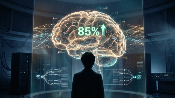


Scientists from the University of Surrey in the UK have developed an innovative artificial intelligence (AI) tool capable of predicting what a person’s knee X-ray will look like in one year, marking a significant advancement in tracking the progression of osteoarthritis. This degenerative joint disorder currently affects more than 500 million people worldwide and is the leading cause of disability among older adults.
By offering both a visual forecast and a risk score, this new technology provides doctors and patients with a more comprehensive understanding of the disease. The AI tool is faster and more interpretable than previous systems, and researchers are optimistic that it could soon be adapted to predict other health conditions, including lung and heart diseases.
The research was recently showcased at the International Conference on Medical Image Computing and Computer Assisted Intervention (MICCAI 2025). It describes a robust AI model that generates realistic “future” X-rays while also estimating disease progression through a personalized risk score. This dual output offers clinicians and patients a visual roadmap of how osteoarthritis may evolve over time.
Trained on nearly 50,000 knee X-rays from approximately 5,000 patients, this model represents one of the largest datasets of its kind. It boasts the ability to predict disease progression around nine times faster than similar tools, operating with enhanced efficiency and accuracy.
At the heart of this advanced system lies a generative model known as a diffusion model. This technology not only creates a “future” version of a patient’s X-ray but also identifies 16 critical points in the joint, highlighting areas that are being monitored for potential changes. This feature promotes transparency, allowing clinicians to see precisely which parts of the knee the AI is tracking, ultimately helping to build confidence in its predictions.
David Butler, the study’s lead author, emphasized the importance of this approach: “We’re used to medical AI tools that give a number or a prediction, but not much explanation. Our system not only predicts the likelihood of your knee getting worse — it actually shows you a realistic image of what that future knee could look like.” He added, “Seeing the two X-rays side by side — one from today and one for next year — is a powerful motivator. It helps doctors act sooner and gives patients a clearer picture of why sticking to their treatment plan or making lifestyle changes really matters. We think this can be a turning point in how we communicate risk and improve osteoarthritic knee care and other related conditions.”
The potential applications of similar AI tools extend beyond osteoarthritis. Researchers are exploring the capacity of such technologies to predict lung damage in smokers or track the progression of heart disease, providing a similar level of visual insight and early warning for these conditions. Efforts are underway to collaborate with healthcare providers to bring this technology into everyday clinical practice.
This enhanced visibility can significantly aid clinicians in identifying high-risk patients sooner, allowing for personalized care that has not been feasible until now. The research findings are published in the journal Medical Image Computing and Computer Assisted Intervention, titled “Risk Estimation of Knee Osteoarthritis Progression via Predictive Multi-task Modelling from Efficient Diffusion Model Using X-Ray Images.”
See also Deep Learning Transforms Kidney Transplant Success Rates by Enhancing Outcome Predictions
 CMU-Q Scholar Urges Arabic AI Research to Address Inclusion Gap for 400M Speakers
 EU AI Act Fuels $202B Growth in European AI Market by 2033, Driven by Horizon 2020
 Periodic Labs Aims to Create Room-Temperature Superconductor Using AI Innovations
 Engineers Accelerate AI & Machine Learning Breakthroughs in Health, Energy, and Manufacturing