


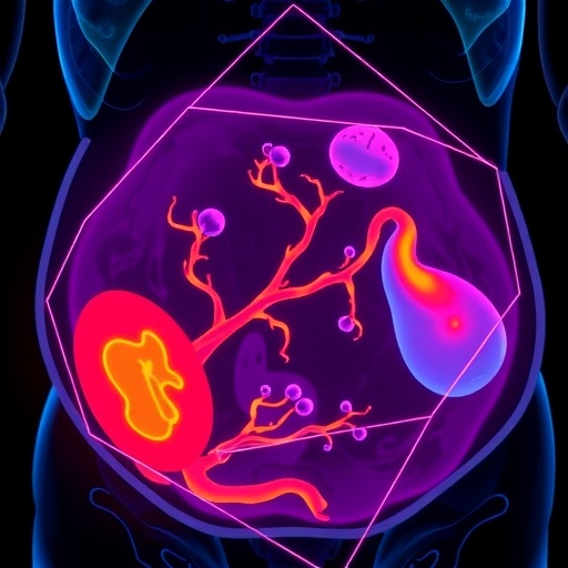
In a significant advancement for maternal-fetal medicine, researchers at Baylor College of Medicine have introduced a novel artificial intelligence (AI) model capable of accurately detecting placenta accreta spectrum (PAS) prior to delivery. PAS, a serious pregnancy complication characterized by abnormal placental adherence to the uterine wall, has long posed diagnostic challenges, contributing to maternal mortality and morbidity worldwide. This pioneering AI-driven diagnostic tool, unveiled at the 2026 Society for Maternal-Fetal Medicine (SMFM) Pregnancy Meeting™, represents a transformative leap toward early identification and intervention in high-risk pregnancies.
Placenta accreta spectrum includes conditions where the placenta invades the uterine wall to varying extents, often associated with previous uterine surgeries like cesarean deliveries. The rising prevalence of PAS in the United States, partly due to increased cesarean rates, has heightened the urgency for reliable screening solutions. Traditional diagnostic methods largely depend on risk factor assessments and sonographic evaluations, yet these approaches are plagued by limitations, such as operator dependence and the risk of inconclusive ultrasound results. As a consequence, nearly half of PAS cases remain undiagnosed until delivery, when severe complications like catastrophic hemorrhage can occur.
To address these diagnostic hurdles, the Baylor research team developed an innovative AI algorithm that analyzes two-dimensional (2D) obstetric ultrasound images with remarkable accuracy. The retrospective study evaluated ultrasound images from 113 pregnant patients deemed high risk for PAS, all of whom delivered at Texas Children’s Hospital between 2018 and 2025. The ultrasounds, conducted around 31 weeks of gestation—a crucial period for prenatal monitoring—provided the dataset for the AI model. This analytical framework employs deep learning techniques to identify subtle morphological patterns indicative of placental invasion that could escape human detection.
The study’s findings were compelling: the AI model achieved perfect sensitivity, successfully identifying every confirmed case of PAS within the cohort. Despite generating two false positive results, it notably refrained from any false negatives, highlighting its potential as a highly reliable screening tool. This degree of diagnostic accuracy could enable obstetricians to better prepare for complicated deliveries, potentially reducing the risks of severe maternal hemorrhage, multi-organ failure, and mortality associated with undiagnosed PAS.
The AI model’s effectiveness lies in its ability to process complex imaging data beyond traditional visual analysis. By harnessing extensive pixel-level information and training on annotated datasets, the algorithm can differentiate between normal placental attachment and pathological adherence. This transcends the variability inherent in human interpretation, offering a standardized and replicable diagnostic resource. Furthermore, integrating AI into obstetric ultrasound workflows could democratize expertise, delivering essential decision support in areas with limited access to specialized maternal-fetal medicine consultations.
Dr. Alexandra L. Hammerquist, a maternal-fetal medicine fellow and lead researcher, emphasized the model’s clinical implications. “Our team is very excited about the potential clinical implications of this model for accurate and timely diagnosis of PAS. We are hopeful that its use as a screening tool will help decrease PAS-related maternal morbidity and mortality,” she stated. This AI-assisted diagnostic approach not only aims to improve the detection of PAS but also facilitates personalized prenatal care pathways, allowing for tailored surveillance intensity and delivery planning.
The retrospective design of the study utilized a comprehensive dataset accumulated over nearly seven years, reflecting real-world clinical variability. Inclusion criteria focused on pregnancies considered high risk due to clinical or obstetric history, making the findings particularly relevant for targeted screening strategies. The choice of 2D ultrasound, rather than more advanced imaging techniques, enhances the applicability of this AI tool, as 2D ultrasound remains the standard imaging method globally due to its accessibility and cost-effectiveness.
Although the presence of two false positives suggests areas for improvement, the lack of false negatives is crucial from a clinical safety standpoint, ensuring that cases of PAS do not go unnoticed. Future prospective studies will be vital for validating these promising findings, assessing the AI model’s efficacy across diverse populations and evaluating its integration into clinical practices. Moreover, exploring real-time applications during ultrasound acquisitions represents an exciting frontier, potentially enabling instant diagnostic support.
The implications of this research extend beyond PAS, underscoring the broader potential for AI to transform prenatal diagnostics. By augmenting human expertise with machine learning capabilities, clinicians can detect subtle pathological features that might otherwise remain unseen, enhancing early detection of various pregnancy complications. This study marks a significant step toward precision obstetrics, where data-driven insights guide optimized therapeutic decisions, ultimately improving outcomes for both mothers and their babies.
This groundbreaking AI model has garnered substantial attention in anticipation of its detailed presentation in oral abstract #39 titled “AI-based ultrasound screening for early, accurate identification of placenta accreta spectrum,” set for publication in the February 2026 issue of Pregnancy, the official peer-reviewed journal of the Society for Maternal-Fetal Medicine. The research symbolizes hope for maternal health, aiming to alleviate the devastating consequences of undiagnosed PAS and establish a new benchmark for diagnostic accuracy in high-risk obstetrics.
As global cesarean delivery rates continue to rise alongside increasing PAS incidence, timely and accurate diagnosis becomes paramount. The integration of AI into obstetric practice, as demonstrated by this study, underscores a future in which technology not only supports but also fundamentally transforms clinical paradigms. Innovations like the Baylor College model present a compelling vision of safer pregnancies through enhanced early detection.
The Society for Maternal-Fetal Medicine, representing over 6,500 specialists focused on managing high-risk pregnancies, highlights the critical need for advancements in this realm by promoting research, education, and advocacy. This AI-based screening breakthrough stands at the crossroads of clinical necessity and technological innovation, poised to deliver significant benefits for maternal and fetal health worldwide.
See also MIT’s J-PAL Launches Project AI Evidence to Evaluate AI Solutions Against Poverty
 World Bank Study Reveals Hierarchy’s Role in AI Research Dissemination among 6,000 Employees
 AI Transforms Alzheimer’s Drug Discovery with New Collaborative Approaches and Data Insights
 Improved Convergence Rates for Federated Variational Inequalities with LIPPAX Algorithm
 Cisco Reports Q2 FY26: $15.3B Revenue Driven by AI Infrastructure Orders