


An artificial intelligence developed by researchers at Kobe University has demonstrated the ability to diagnose acromegaly, a rare endocrinological condition, through the analysis of photographs of the back of the hand and a clenched fist. This privacy-centric breakthrough, which has the potential to enhance healthcare referral systems and address health disparities, was published in the Journal of Clinical Endocrinology & Metabolism.
Acromegaly, primarily affecting middle-aged individuals, is characterized by abnormal growth of the hands and feet, changes in facial features, and various complications related to bone and organ growth. Caused by an overproduction of growth hormone, the condition progresses slowly over decades, often leading to life-threatening complications and a decrease in life expectancy by approximately 10 years. Dr. Fukuoka Hidenori, an endocrinologist at Kobe University, noted that the slow progression of the disease often results in diagnostic delays of up to a decade.
Current AI models for detecting acromegaly have typically relied on facial images, raising privacy concerns. Dr. Ohmachi Yuka, a graduate student at Kobe University, explained that the research team decided to focus on the hands, an area frequently examined alongside the face in clinical diagnostics. By using images exclusively of the back of the hand and the clenched fist, the team ensured greater privacy while still capturing significant changes associated with acromegaly. This approach facilitated the involvement of 725 patients across 15 medical facilities in Japan, who collectively contributed over 11,000 images to train and validate the AI model.
The published results indicate that the AI model achieves high sensitivity and specificity in diagnosing acromegaly, even surpassing the accuracy of experienced endocrinologists who assessed the same photographs. “I was surprised that the diagnostic accuracy reached such a high level using only photographs of the back of the hand and the clenched fist,” said Ohmachi. “What struck me as particularly significant was achieving this level of performance without facial features, which makes this approach a great deal more practical for disease screening.”
Looking ahead, the research team aims to extend their model to other conditions identifiable through similar photographic methods, such as rheumatoid arthritis, anemia, and finger clubbing. Ohmachi suggested, “This result could be the entry point for expanding the potential of medical AI.” While hand images are not a standalone diagnostic tool in medical practice, the Kobe University team sees the model as a means to complement clinical expertise, mitigate diagnostic errors, and facilitate earlier interventions.
Dr. Fukuoka emphasized the broader implications of their work, suggesting that further development of this technology could contribute to establishing a medical infrastructure during comprehensive health check-ups. This could improve the referral of suspected cases of hand-related disorders to specialists and provide non-specialist physicians in regional healthcare settings with necessary support, ultimately narrowing healthcare disparities.
This research was funded by the Hyogo Foundation for Science Technology in collaboration with several leading institutions, including Fukuoka University, Hyogo Medical University, Nagoya University, and others.
For more details, refer to the study: DOI: 10.1210/clinem/dgag027.
See also Perplexity Launches Perplexity Computer, a Multi-Model AI Research Assistant for Complex Tasks
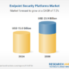
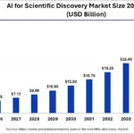 AI for Scientific Discovery Market Projected to Reach $34.78B by 2035 with 21.90% CAGR
 Study Reveals Diverse Pre-Training Data Boosts Retinal AI Model Performance and Equity
 AI Boosts Research Output by 3x but Reduces Topic Diversity, Study Finds