


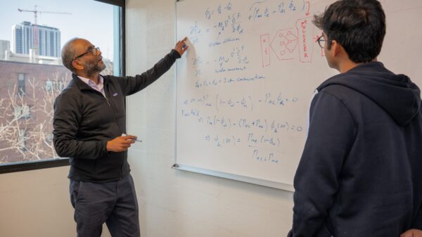


In an era where artificial intelligence is increasingly infiltrating various sectors, its potential application in orthopedics is gaining traction. Researchers, led by Michael A. David, PhD, an instructor at the University of Colorado Anschutz School of Medicine, are exploring how machine learning (ML) tools can help predict bone fractures and classify their types, aiming to enhance patient care.
David emphasizes that while these advanced tools are not yet commonplace in clinical settings, their use in research is expanding. “The appeal is that these tools can take a large amount of data and efficiently make sense of it with minimal human input,” he says. His work integrates ML with disciplines such as spatial histopathology, transcriptomics, and medical imaging to address post-traumatic and idiopathic joint conditions, including contracture, osteoarthritis, osteoporosis, and tendon ruptures.
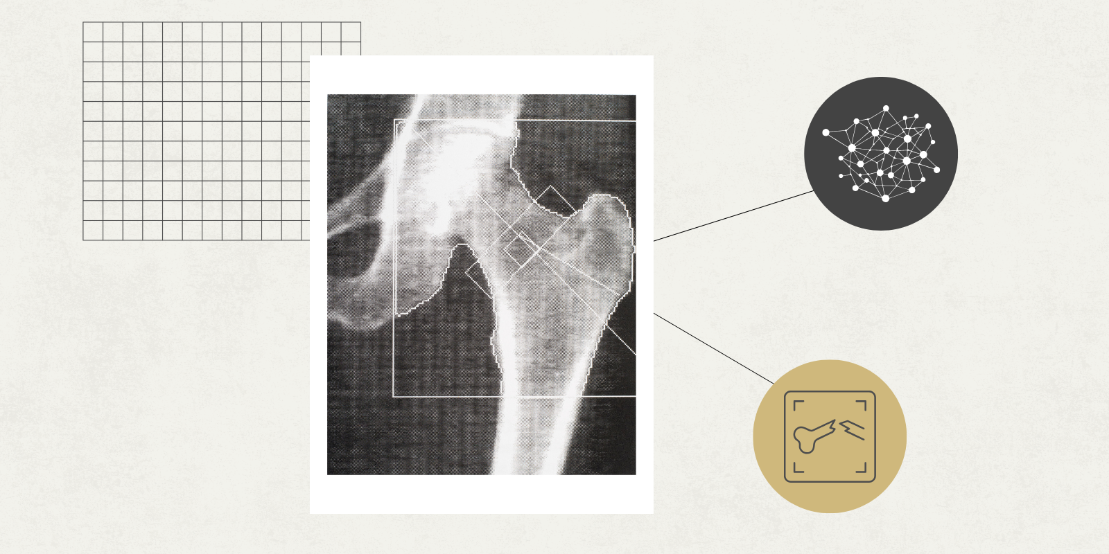
In a recent review published in Bone Reports, David discusses the potential for ML to facilitate agnostic, automated analysis, which he believes could accelerate discovery and enhance clinical bone diagnosis and treatment monitoring. One prominent application of ML is in performing segmentation, which involves dividing digital bone images into distinct types for faster processing. This capability is particularly beneficial in orthopedic research, where significant amounts of data are necessary for effective analysis.
“Segmenting bone data is the most laborious, time-consuming task,” states David, reflecting on his experiences as a graduate student. “That’s one area where ML excels in terms of accelerating the pace of discovery.” By automating this process, ML not only speeds up the workflow but also extracts vital quantitative data from bone images, providing researchers with clearer insights.
However, David notes that not all bone images contain uniform information, necessitating human expertise to ensure the effectiveness of ML applications. His review focuses primarily on two imaging techniques commonly used to assess bone structure: micro-computed tomography (microCT) and high-resolution peripheral quantitative computed tomography (HRpQCT). He reports that ML has primarily been utilized to enhance image resolution, automate segmentation, reduce experimental variables, cluster data, and assist in predicting and classifying bone health and fracture risk.
While these technological advancements are promising, David is quick to clarify that ML is unlikely to replace radiologists. Instead, he envisions these methods as clinical tools that will support healthcare providers in making optimal decisions for their patients. Additionally, by clustering patient data, ML can reveal insights into subtypes or stages of diseases like osteoporosis, ultimately benefiting patient outcomes.
“With ML, you can combine many types of data, like bone imaging, genetics, and even sleep cycle information, to see whether subgroups form,” David explains. He believes that substantial progress can be made in this area, showcasing the transformative potential of integrating diverse data sources.
As ML becomes more prevalent, David stresses the importance of researchers gaining a comprehensive understanding of these technologies. “Although significant information is available in the public domain, particularly with the rise of large language ML models such as Deepseek, ChatGPT, Claude, and Gemini, the sheer breadth of tools and metrics creates a steep learning curve,” he notes. This complexity can act as a barrier for bone researchers aiming to adopt ML methodologies in their work.
To address this challenge, David authored the review paper and developed reproducible code for foundational ML programming. He also created SciNetX, a scientometric and bibliometric analytical pipeline and visualization software. “SciNetX was born from the need to structure and illustrate how all the research papers fit together,” he says. The software allows users to input various types of information, such as paper subjects, keywords, and authors, generating visual networks that illuminate connections among different research topics and investigators.
“It’s basically a map of the field,” David adds, highlighting its utility for new researchers. Although initially developed for orthopedic applications, SciNetX has broader applicability across various medical research areas and beyond. He is currently collaborating with CU Innovations to license the software for wider use.
Ultimately, David envisions a future where human and computer interactions are symbiotic, fostering a more inclusive environment for those interested in incorporating ML in digital bone imaging. “The whole idea is to invite more people into this world,” he concludes. His efforts aim to empower researchers to approach this field ready to learn, thereby accelerating advancements in orthopedic research and patient care.
See also OpenAI’s GPT-5 Conducts 36,000 Experiments, Highlighting AI’s Risks in Biology
 Evo 2 AI Model Predicts Genetic Disease-Causing Mutations from 128,000 Genomes
 New Deep Learning Framework Achieves 36.8% Boost in Drone-Based Vehicle ReID Accuracy
 Bessent Declares Anthropic’s Mythos a Game-Changer in US-China AI Competition